Published on: September 23, 2019 | Updated on: August 29, 2024
Our hips are a vital aspect of movement in day-to-day activities. They are used in almost all activities, from running to walking, sitting down, and even wiggling toes.
When the hip becomes injured it can affect all of these aspects of daily life. Hip dysplasia affects around 1 out of every 1,000 infants. The disease is more common in girls and firstborn children. It can happen in either hip, although the left side is more prevalent.
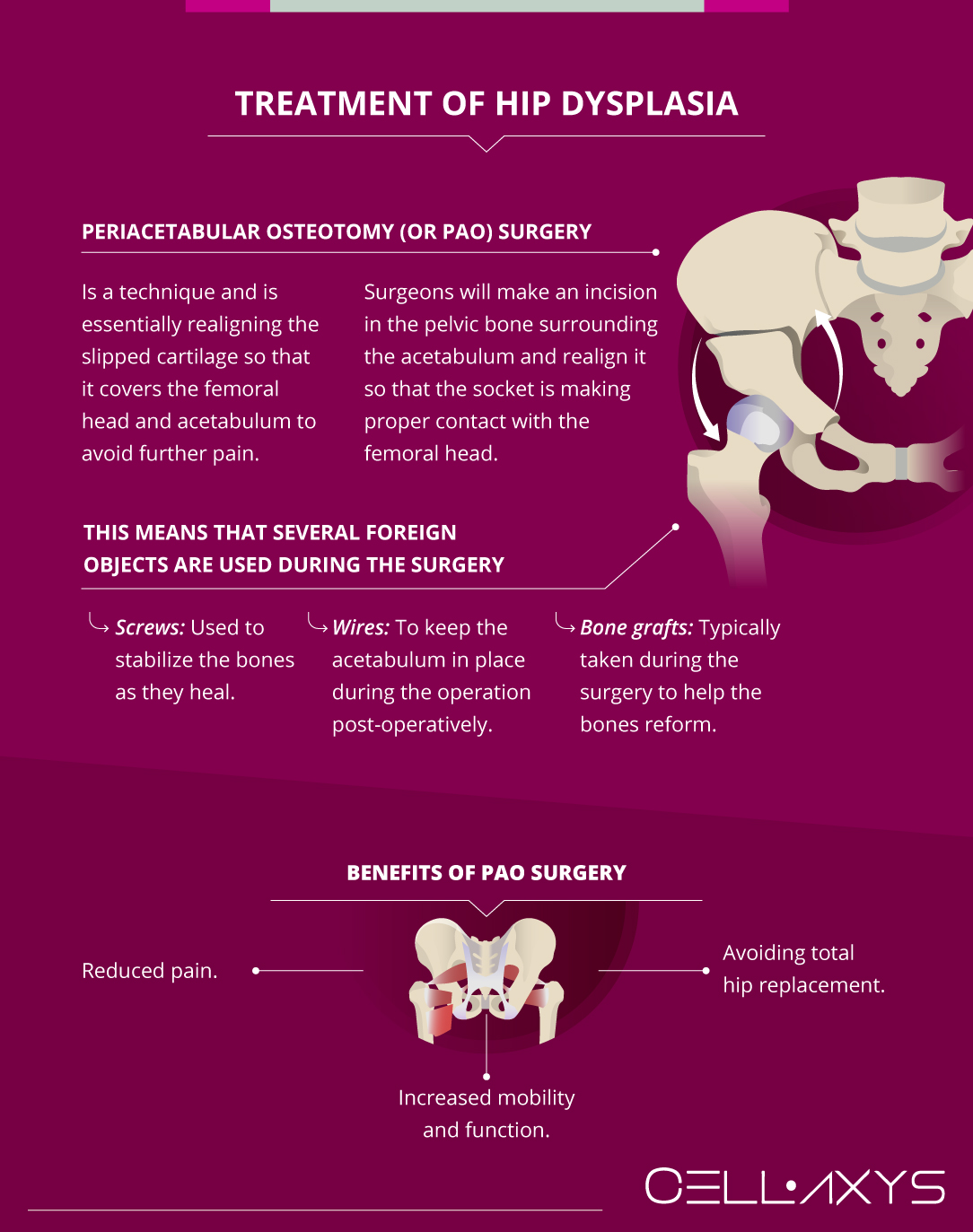
The most popular option for the treatment of hip dysplasia is periacetabular osteotomy (or PAO) surgery, an elective surgery that corrects the damage done from hip dysplasia.
Anatomy of the Hip Joint
The hip joint is a ball-and-socket joint, meaning that it functions by using a round, ball-like portion that moves around in a socket. Due to its shape, this type of joint has a high range of motion (similar to the shoulder joint) but is heavily dependent on proper cartilage formation. There are two bones that comprise the joint and two types of cartilage that exist between the ball and socket:
- Femoral head: the round head of the thigh bone is what makes up the ball part of the joint. The femoral head fits into the acetabulum.
- Acetabulum: the socket part of the joint, located in the pelvic bone.
- Articular cartilage: covers the femoral head and acetabulum to allow for movement within the joint. Its location prevents the two bones from rubbing against one another.
- Labrum: attached to the articular cartilage and allows for the joint to be stabilized by expanding the socket.
What is Hip Dysplasia?
In hip dysplasia, the femoral head is not entirely covered with cartilage as it should be. The acetabulum may also be more shallow than it should be, which can lead to the femoral head being pushed out of the socket over time.
The hip joint might become partly or totally dislocated as a result of this. The lack of cartilage can lead to bones rubbing against one another, which causes pain and a limited range of motion.
Signs and Symptoms of Hip Dysplasia
Signs of hip dysplasia can occur during infancy all the way to old age. In some infants, it can be easier to diagnose because there may be one leg that is longer than the other or less mobility in one of the hips. As a person ages, the signs may become more prominent.
The early signs of hip dysplasia that occur if not diagnosed in infancy are:
- Walking with a limp
- Pain in the groin
- Pain during certain activities such as sitting, walking, or exercise
- Pain during rest or at night
- A feeling or sound similar to popping or locking
The pain associated with hip dysplasia is typically felt deep in the groin area but can be felt all over the general hip area. The pain can begin as a dull ache or can feel like a sharp, stabbing pain. It can be experienced during certain activities or can be chronic. This pain may begin at any point in life.
Causes
Hip dysplasia is slightly more common in women than men and is typically a condition that exists from birth.
If left untreated, hip dysplasia can lead to osteoarthritis later in life. Arthritis is characterized by a degradation of cartilage within joints that causes bones to rub against one another. If an individual is already suffering from a lack of sufficient cartilage in the hip joint, the body’s natural degradation of cartilage can become osteoarthritis.
The nature of hip dysplasia makes room for other injuries to occur. When the joint is generally misaligned, the tendons and muscles surrounding it can become overworked or stressed as they attempt to correct the abnormality.
This can lead to a tear in the labrum or surrounding muscles. The symptoms of these injuries are often similar to those of hip dysplasia, so it is important that they are diagnosed properly.
Diagnosing Hip Dysplasia
In non-infant patients who have hip dysplasia, doctors will use several tests to determine the cause of hip pain. These can include:
- Physical examination: doctors will watch a patient walk in an attempt to observe a limp. They also might observe the patient’s range of motion in the hip or lightly touch areas that may be causing pain. These tests are a simple way to target the location of the pain, though they do not necessarily determine the cause of pain.
- X-ray imaging: an X-ray can determine the location of bones that may be misaligned due to hip dysplasia. The lack of cartilage to separate the bones sometimes causes the femoral head to move up and out of the acetabulum, and this can be observed using X-Ray technology.
- MRI imaging: an MRI may be ordered to capture an image of the soft tissue surrounding the joint, including the malformed cartilage. This type of imaging is also able to capture images of tears in muscles, tendons, or cartilage in the area.
- CT scan: this type of imaging is able to create a comprehensive image of the bones and soft tissue, allowing doctors to gather a full image of the injury.
The difficulty in diagnosing hip dysplasia arises from the myriad of other injuries that may be happening in the area, some of which may have been caused by dysplasia. For some patients, it takes visiting more than one doctor and several tests in order to properly diagnose the disorder.
It is vital that it is diagnosed properly so that it can receive proper treatment – overlooking a potential cause of injury could lead to unnecessary surgery, increased pain over time if an issue is left untreated, or increased damage.
Treatment of Hip Dysplasia

Unlike other hip injuries, treatment of hip dysplasia almost always results in a surgery called Periacetabular Osteotomy (or PAO) surgery. In the case of labral tears or hip impingement, treatment typically begins with rest and may eventually lead to corrective surgery, but the body is capable of healing itself over time with these types of injuries.
With hip dysplasia, the lack of cartilage that is causing pain is not capable of healing itself in such a way, leading to PAO surgery.
There is another form of surgery called arthroscopy that may be performed to correct any tears in the cartilage or surrounding soft tissues. Arthroscopy is performed using a tiny camera and specially designed instruments that are inserted into the joint to correct tears in soft tissue.
Due to its minimally invasive nature, this type of surgery does not correct the cartilage in the hip joint if it has receded and is not capable of realigning the bones for optimal function.
PAO surgery is performed with a slightly more invasive technique. Surgeons will make an incision in the pelvic bone surrounding the acetabulum and realign it so that the socket is making proper contact with the femoral head.
This surgery involves not only cutting into bone but also realigning it. This means that several foreign objects are used during the surgery. These foreign objects include:
- Screws: used to stabilize the bones as they heal
- Wires: to keep the acetabulum in place during the operation
- Bone grafts: typically taken during the surgery to help the bones reform post-operatively.
The number of tools used, locations of incisions, and alignment of the bone is all done specific to the patient’s needs, meaning that each surgery is somewhat unique. The aforementioned imaging procedures are used to determine a patient’s specific needs.
If the cartilage is not yet too damaged, this surgery provides the joint a return to proper function. Some of the benefits of PAO surgery include:
- Reduced pain
- Increased mobility and function
- Avoiding total hip replacement
Total hip replacement is typically considered when a patient is also suffering from some form of osteoarthritis and is seen as a last resort to treating hip pain. If hip dysplasia is the cause of this pain and osteoarthritis, PAO surgery will be an option long before a total hip replacement is considered.
Depending on the extent of the injury, some surgeons may also elect to correct damage to the surrounding tissue while they are undergoing PAO surgery.
All surgeries come with some risks; mainly there are risks of anesthetic complications or rejection, which may include pneumonia, blood clots, stroke, or even death. There is also a risk of infection from the environment.
Recovery From PAO Surgery
It is important that patients who have undergone PAO surgery follow their doctor’s recovery plan directly. All recoveries involve resting the joint as it heals at first. After a couple of weeks, physical therapy can begin.
Physical therapy allows a patient to restrengthen the muscles surrounding the joint so that it can increase support and mobility. The ability to walk using an aid such as crutches is typically possible within a few days after the surgery but should be closely monitored and limited.
The success of the surgery is dependent on the patient’s ability to follow a recovery procedure that has been laid out for them.
Each recovery process is different from the next depending on the patient’s anatomy, physical activity level prior to the procedure, and what surgical tactics were used. It is also possible to speed up the recovery process using regenerative therapies.
How Regenerative Medicine Can Boost PAO Surgery Recovery Time?
The constantly growing field of regenerative medicine has offered hope for a myriad of conditions. They work well on soft-tissue injuries, meaning that they are strong candidates for helping the hip joint heal after PAO surgery. The two major types of regenerative therapy on the market are:
- Autologous Stem Cell & Cell Based Procedure: this form of therapy begins with taking a sample from a patient’s fat cellsor bone marrow. This tissue is then processed to isolate the autologous mesenchymal stem cells and other cells, which are then transplanted into the area of pain. These cells are able to engage other cells in the body which contain healing properties. With an increase in healing properties available, injury, damage, and surgical locations can heal better. Cell Based Procedures can be use for both recovery from surgery and for continued post-operative pain.
- Platelet-Rich Plasma (PRP) Therapy: the PRP process starts with a simple blood draw from the patient. The blood is then placed in a centrifuge to separate the platelets from the plasma. Platelets contain growth factors and proteins that aid in the healing process. These are then reinjected to the injured area.
Both of these procedures use ultrasound or fluoroscopy imaging technology to locate the exact location of the injury/pain where the transplant will occur. With PAO surgery, doctors may choose to use X-ray imaging to guide them to the surgical location where they will proceed with the transplant.
Some surgeons will also elect to use a certain type of string during surgery that is infused with stem cells or platelets. This allows them to interact with the injury site more intimately, which in turn increases their potential to heal the surgical site.
Regenerative therapy can help with recovery significantly, as be beneficial to post-op chronic pain if present.
Sources
Footnotes
- Jashi RE, Gustafson MB, Jakobsen MB, Lautrup C, Hertz JM, Søballe K, Mechlenburg I. The association between gender and familial prevalence of hip dysplasia in Danish patients. Hip International. 2017;27(3):299-304.
- Loder RT, Skopelja EN. The epidemiology and demographics of hip dysplasia. International Scholarly Research Notices. 2011;2011.
- Schroeder C, Zavala L, Opstedal L, Becker J. Recovery of Lower Extremity Function in the Initial Year Following Periacetabular Osteotomy: A Single Subject Analysis. Physiotherapy Theory and Practice. 2022;38(9):1233-44.
- Schroeder CM. Rehabilitation outcomes following a periacetabular osteotomy (PAO): a case study. Doctoral dissertation, Montana State University-Bozeman, College of Education, Health & Human Development. 2021.
- Petrie JR, Novais EN, An TW, Schoenecker PL, Zaltz I, Kim YJ, Millis MB, Beaule PE, Sierra RJ, Trousdale RT, Sucato DJ. What is the impact of periacetabular osteotomy surgery on patient function and activity levels?. The Journal of Arthroplasty. 2020;35(6):S113-8.
- Ruzbarsky JJ, Comfort SM, Rutledge JC, Shelton TJ, Day HK, Dornan GJ, Matta JM, Philippon MJ. Improved functional outcomes of combined hip arthroscopy and periacetabular osteotomy at minimum 2-year follow-up. Arthroscopy: The Journal of Arthroscopic & Related Surgery. 2024;40(2):352-8.
References
- Periacetabular Osteotomy: An Overview. HSS. Accessed 2/23/2024.
- Periacetabular Osteotomy (PAO). University of Utah Health. Accessed 2/23/2024.
- What to Expect If You or Your Child Need Periacetabular Osteotomy. Healthline. Accessed 2/23/2024.
- Arthritis. Mayo Clinic. Accessed 2/23/2024.
- Arthroscopy. OrthoInfo. Accessed 2/23/2024.
CELLAXYS does not offer Stem Cell Therapy as a cure for any medical condition. No statements or treatments presented by Cellaxys have been evaluated or approved by the Food and Drug Administration (FDA). This site contains no medical advice. All statements and opinions are provided for educational and informational purposes only.
Dr Pouya Mohajer
Author
Pouya Mohajer, M.D. is the Director of Spine and Interventional Medicine for CELLAXYS: Age, Regenerative, and Interventional Medicine Centers. He has over 20 years of experience in pain management, perioperative medicine, and anesthesiology. Dr. Mohajer founded and is the Medical Director of Southern Nevada Pain Specialists and PRIMMED Clinics. He has dedicated his career to surgical innovation and scientific advancement. More about the doctor on this page.
Dr Pejman Bady
Contributor
Dr. Pejman Bady began his career over 20 years ago in Family/Emergency Medicine, working in fast-paced emergency departments in Nevada and Kansas. He has served the people of Las Vegas as a physician for over two decades. Throughout this time, he has been met with much acclaim and is now the head of Emergency Medical Services in Nye County, Nevada. More about the doctor on this page.


