Published on: February 21, 2024 | Updated on: September 4, 2024
Magnetic Resonance Imaging (MRI) is a powerful method that gives high-quality pictures of bones, tendons, cartilage, and nerves without radiation. It is considered a better alternative to X-rays that only capture bones. Doctors use MRI to see different detailed views of the neck, called cervical spine MRI.
MRI helps healthcare providers identify the exact location of a given structure in case of a disease. For instance, disc herniation requires the doctor to know exactly where the issue is and whether its symptoms are widespread.
A normal MRI captures cervical bones, discs, facet joints, central canal, neural foramen, muscles, and tendons. Meanwhile, abnormal cervical spine MRI shows cervical spondylosis, disc injuries, bone spurs, facet injuries, subluxation, etc.
Let’s take deeper insights into normal vs abnormal cervical spine MRI.
What Is a Cervical Spine MRI?

What Is a Cervical Spine MRI?
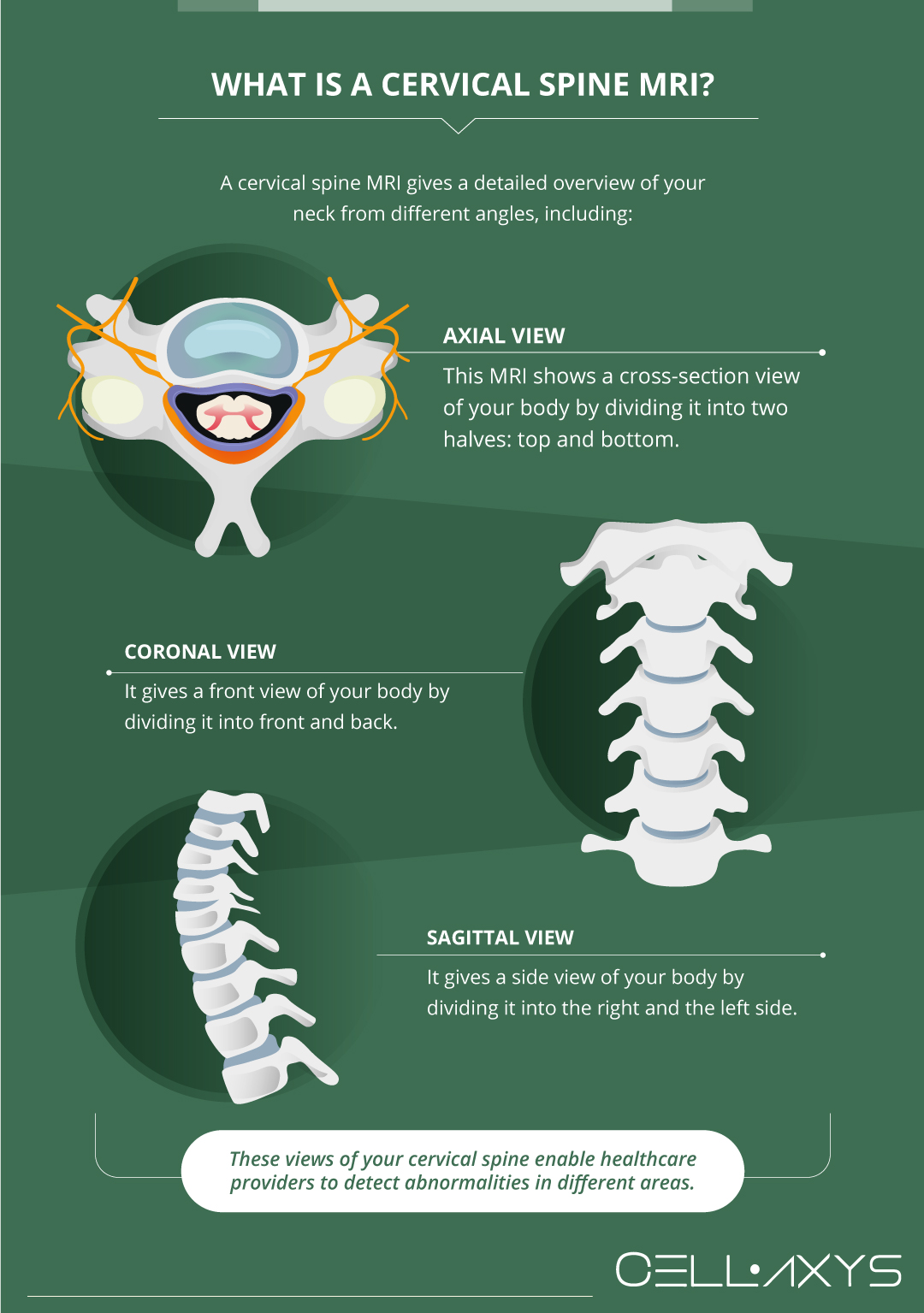
A cervical spine MRI gives a detailed overview of your neck from different angles, including:
- Axial View. This MRI shows a cross-section view of your body by dividing it into two halves: top and bottom.
- Coronal View. It gives a front view of your body by dividing it into front and back.
- Sagittal View. It gives a side view of your body by dividing it into the right and the left side.
These views of your cervical spine enable healthcare providers to detect abnormalities in different areas.
What Is a Cervical Spine MRI Used for?
Your doctor may recommend a cervical spine MRI for many reasons, such as:
- Severe neck pain
- Arm pain that persists after conventional treatments
- Neck trauma
- Infection
- Scoliosis (Curvature)
- Headaches
- Tingling in the arm
- Spinal defects
Normal Cervical MRI: What Does It Look Like?
The cervical spine comprises various parts, each working collaboratively to keep your neck intact and fully mobile. A normal cervical spine MRI shows the imagery of the following parts:
Cervical Bodies and Discs
The neck consists of seven blocks called vertebral bodies, numbered from C1 to C7 from top to bottom. The “C” refers to the cervical spine. Each vertebral column is separated by a cushion from the next body, known as a disc. The disc keeps the neck intact and ensures its proper functioning.
Muscles & Tendons
The neck has lots of large and small muscles and tendons in the neck. The tendons are connective tissues connecting the muscles to the bones. Both play a major role in the spine’s stability and mobility.
A normal cervical spine MRI thoroughly scans the neck’s muscles and tendons.
Central Canal
The central canal runs across the spine, including the spinal cord and fluid. A normal MRI can give a detailed scan of the central canal.
Facet Joints
Facet joints are paired, cartilage-lined joints present in the back area of the neck. There are two types of facet joints — right and left — each located at every cervical level. They are named after the vertebral bodies they are present close to.
For example, the C3 and C4 neck bones make up the C3/4 facet joint. These joints provide support and stability to the neck and manage its range of motion.
Neural Foramen
The spine consists of the neural foramen, the path from which the spinal nerve roots exit the spinal column. Each spinal level has a neural foramen; they are also present on the right and left sides.
Abnormal Cervical Spine MRI: What Does It Scan?
The abnormal cervical spine MRI findings include the scans of:
Cervical Spondylosis
Cervical spondylosis is a broad term used for degenerative arthritis of the neck. The condition can get worse with a lack of treatment.
Disc Injuries
The cervical discs absorb trauma and shock to the neck, but they are susceptible to various injuries. The cervical spine MRI can easily detect any deformity to the disc, called disc protrusion, extrusion, and herniation.
Subluxation
The cervical bones are aligned to make up the cervical spine. This arrangement enables optimal neck functionality, but any trauma can affect it. When one vertebral bone moves forward, it is called Anterolisthesis.
“Listhesis” means slippage, and “anterior” refers to forward. When the vertebral body slips backward, the condition is called Retrolisthesis. Both these issues are collectively called Subluxation, leading to intense pain in the neck.
An abnormal cervical spine MRI can detect the subluxation.
Bone Spurs
Bone spurs are small, large, irregular, or smooth growth on the bones due to micro instability. The body produces these bony projections to ensure stability, but they can pressurize your nerves and narrow the central canal and the neural foramen. The MRI scan can easily detect bone spurs.
Facet Injuries
Any injury to the facet joints present in the back of the neck can be identified through the MRI scan. The degeneration of the joint can cause pain and limited mobility of the neck, which worsen over time.
Central Canal Stenosis
When the canal becomes narrowed, it refers to stenosis. It is diagnosed based on the severity of the condition. The stenosis in the central canal can cause neck pain, mobility issues, and numbness in the arms and hands.
Foraminal Stenosis
The neural foramen (the boney doorway) can become narrow, leading to foraminal stenosis. In this condition, the spinal nerves don’t find the pathway to exit from the spinal canal, thereby becoming compressed or irritated.
The doctor uses MRI to detect these issues in the cervical spine.
The Best Alternatives to Cervical Spine MRIs
The major differences between a normal vs abnormal cervical spine MRI lie in the overall health of the cervical discs, vertebral columns, facet joints, muscles, tendons, and ligaments. However, MRIs don’t evaluate the entire cervical spine, including its stability, health, and gravitational impact.
Depending on your condition, your doctor may opt for the following alternatives to traditional MRI imaging:
- Upright MRI. It evaluates the neck’s stability from different positions when the patient is sitting or standing. You may have to bend a little forward or backward for the MRI to detect cervical instability.
- Digital Motion X-ray. The digital motion X-ray is another effective alternative to determine craniocervical instability. You must move in the side bending, extension, or flexion positions to get the MRI done.
At CELLAXYS, our board-certified surgeons clearly understand the differences between a normal vs abnormal cervical spine MRI. Your MRI report helps us determine the best regenerative treatment for your cervical condition, including cell-based and platelet-rich plasma (PRP) therapies. Connect with us today to book an appointment!
Sources
Footnotes
- Katti G, Ara SA, Shireen A. Magnetic resonance imaging (MRI)–A review. International journal of dental clinics. 2011;3(1):65-70.
- Khoo VS, Dearnaley DP, Finnigan DJ, Padhani A, Tanner SF, Leach MO. Magnetic resonance imaging (MRI): considerations and applications in radiotherapy treatment planning. Radiotherapy and Oncology. 1997;42(1):1-5.
- Rowe L, Steiman I. Anterolisthesis in the cervical spine–spondylolysis. Journal of Manipulative and Physiological Therapeutics. 1987;10(1):11-20.
- Jenis LG, An HS, Gordin R. Foraminal stenosis of the lumbar spine: a review of 65 surgical cases. American Journal of Orthopedics (Belle Mead, NJ). 2001;30(3):205-11.
References
- Magnetic Resonance Imaging (MRI). The Johns Hopkins Hospital. Accessed 8/29/2023.
- Magnetic Resonance Imaging (MRI). National Institute of Biomedical Imaging and Bioengineering (NIBIB). Accessed 8/29/2023.
- Cervical MRI Scan. University of California San Francisco. Accessed 8/29/2023.
- Magnetic Resonance Imaging (MRI): Cervical Spine. The Nemours Foundation. Accessed 8/29/2023.
CELLAXYS does not offer Stem Cell Therapy as a cure for any medical condition. No statements or treatments presented by Cellaxys have been evaluated or approved by the Food and Drug Administration (FDA). This site contains no medical advice. All statements and opinions are provided for educational and informational purposes only.
Dr Pouya Mohajer
Author
Pouya Mohajer, M.D. is the Director of Spine and Interventional Medicine for CELLAXYS: Age, Regenerative, and Interventional Medicine Centers. He has over 20 years of experience in pain management, perioperative medicine, and anesthesiology. Dr. Mohajer founded and is the Medical Director of Southern Nevada Pain Specialists and PRIMMED Clinics. He has dedicated his career to surgical innovation and scientific advancement. More about the doctor on this page.
Dr Pejman Bady
Contributor
Dr. Pejman Bady began his career over 20 years ago in Family/Emergency Medicine, working in fast-paced emergency departments in Nevada and Kansas. He has served the people of Las Vegas as a physician for over two decades. Throughout this time, he has been met with much acclaim and is now the head of Emergency Medical Services in Nye County, Nevada. More about the doctor on this page.


