Published on: September 24, 2019 | Updated on: August 29, 2024
Ulnar-sided wrist pain is difficult to diagnose and even more difficult to treat. Upsetting the fragile biomechanics of the many ulnar side bones of the wrist can lead to grave consequences including pain, reduced range of motion, and loss of grip strength.
Although conventional treatments exist and prove effective in some cases, their effects may be amplified through the use of regenerative therapies.
Do you have Wrist Pain?
Fill out the form below to schedule your FREE virtual consultation
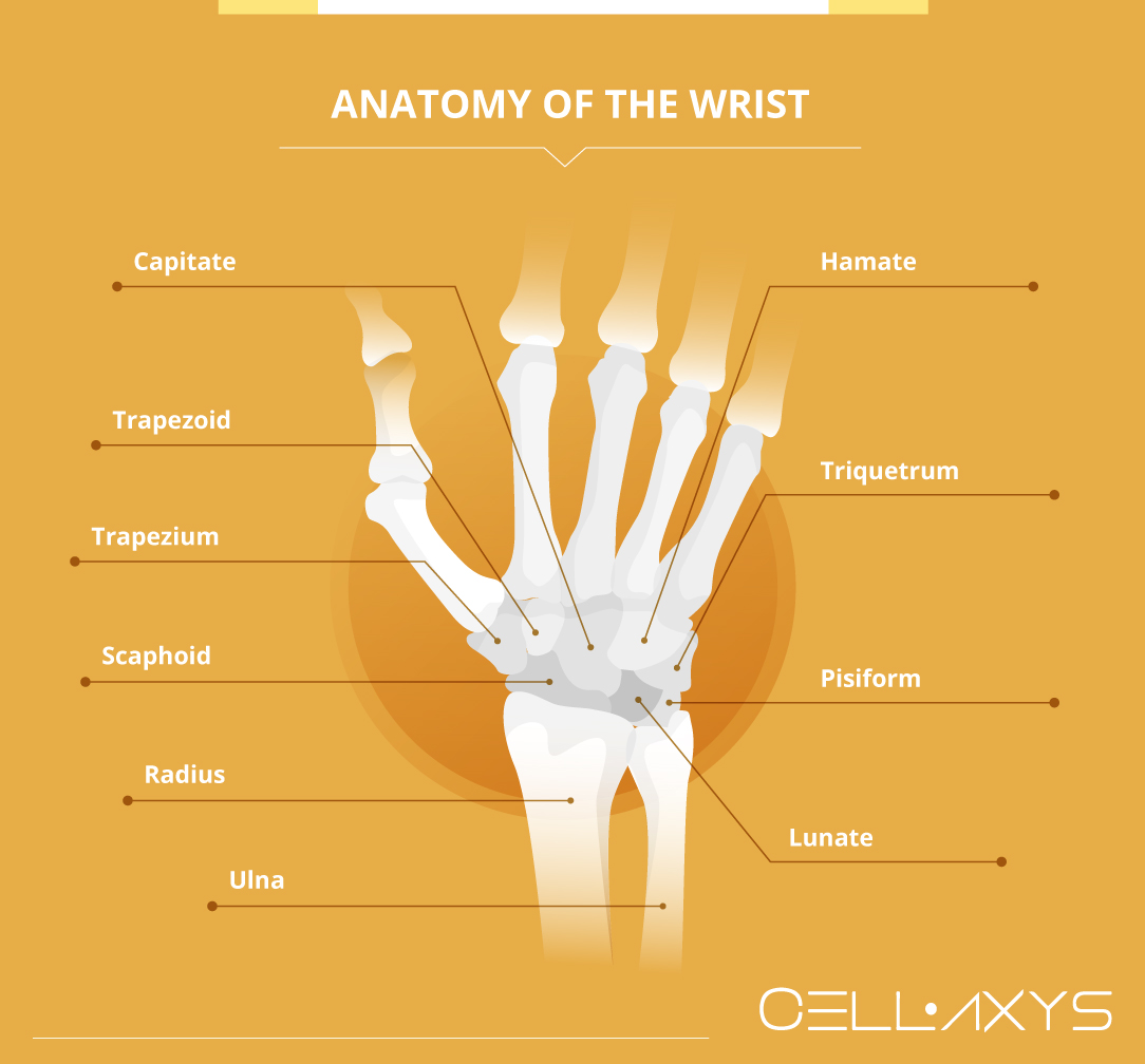
Anatomy of the Wrist

The wrist is composed of a puzzle-like assortment of small bones that have very delicate interactions. To understand the causes of ulnar wrist pain, it is noteworthy to know these bones and their functions:
- Scaphoid
- One of the 2 bones at the base of the wrist. Found between the trapezium and the radius bone of the forearm.
- Covered with cartilage to provide smooth movements between it and 5 other bones in the wrist and forearm.
- Difficult to heal if injured.
- Lunate
- Another of the base bones of the wrist sits directly next to the scaphoid and on top of the radius.
- Rarely injured, but can be involved in wrist dislocations and may rub against the ulna of the forearm if the ulna is too long compared to the radius bone.
- Triquetrum
- Arched above the lunate and under the hamate on the small finger side of the wrist.
- Gives the wrist stability and creates a joint in the wrist with other carpal bones.
- Trapezoid
- Sits above the scaphoid and below the bones of the index finger.
- Uncommonly injured.
- Trapezium
- Sits above the scaphoid and below the thumb.
- It allows for lateral movements of the thumb and provides stability.
- Injures and develops arthritis easily.
- Capitate
- Located under the middle finger, between the trapezoid and the hamate.
- Hamate
- Supports the ring and little fingers.
- Frequently broken.
- An attachment point for the ligament involved in carpal tunnel syndrome.
- Pisiform
- Nestled directly above the triquetrum and lies within a tendon.
- Occasionally breaks or develops arthritis in the joint it makes with the triquetrum.
Also of note are the 2 bones of the forearm:
- Radius
- Sits under the thumb-side of the wrist.
- Ulna
- Sits under the little-finger side of the wrist.
- The root cause of ulnar-sided wrist pain.
A healthy interaction between all of these bones is critical to preventing ulnar-sided wrist pain.
Signs and Symptoms
Ulnar wrist pain can be an indication of various medical conditions. Common signs an individual may have a deeper wrist issue include:
- Ulna (pinkie) sided pain in the wrist.
- Popping or clicking induces pain throughout the base of the hand.
- Pain when gripping or reduction of grip strength.
- Reduced range of motion for the wrist or motion induces a pain response.
- Hand often goes numb or falls asleep
If these signs are a daily occurrence, it may be a sign that the damage is becoming progressively worse and that it may be time to consult a physician.
Causes and Diagnosis
Doctors may have a difficult time identifying the cause of a patient’s pain due to the many individual bones which make up the wrist. Typically, a consultation will involve various range-of-motion exercises and palpation of the wrist by a physician or their assistant.
This examination will most likely be followed by an X-ray, MRI, CT scan, or in rare cases, a micro-invasive procedure using a small guided camera within the wrist.
Some of the most probable causes a doctor will look out for to diagnose a patient’s ulnar-sided wrist pain include:
- Past medical history including wrist and forearm fractures.
- Arthritis of the joint(s) between the wrist bones.
- Ulnar impaction syndrome (caused by an elongated ulna which causes erratic wrist mechanics).
- Inflammation or irritation of the tendons that bend and extend the wrist.
- Triangular fibrocartilage complex (TFCC) (when the connection between the ulna bone and other structures in the wrist is torn by an injury or frayed over time).
- Nerve damage or compression.
Once a doctor understands the root cause of the ulnar-sided wrist pain, they will provide their treatment options. These may include simple physical therapy routines which may be done at home, medication to reduce inflammation or pain response, or, in some cases, an invasive procedure known as ulnar shortening.
Complications of Ulnar Shortening Osteotomy
A healthy interaction between the ulna of the forearm and the bones of the wrist helps ensure an individual has pain-free, normal hand motion. When the ulna pushes on the wrist, or if there has been a past injury to the ulnar-sided wrist bones, normal motions of the hand can induce pain.
Over time, this soreness can weaken your grip, create numbness or pain, and restrict your wrist’s range of motion. Doctors would often prescribe ulnar shortening surgery if these issues become serious and traditional treatment approaches are ineffective.
By reducing the length of the ulna, surgeons can relieve compression of the nerves in the wrist, thereby reducing pain. This shortening is achieved by making incisions in the ulna and grafting the two separate pieces back together using metal plates to hold them in place.
While initial recovery is short, patients typically take at least 4 months before returning to work. Patients are also required to take physical therapy post-surgery to gradually return wrist strength and may require prolonged use of medication to treat post-surgical pain.
In addition to lengthy recovery periods, complications with the mechanics of ulnar shortening surgeries exist. One study which spanned over 10 years reported that over half (51%) of their patients described metalwork irritation from the plates attached in the procedure. Other complications included non-union or misalignment (6.1%), refracture (1.6%), and chronic regional pain (1.6%).
While symptoms did improve in many patients, the risks inherent to ulnar shortening surgery may outweigh the benefits for individuals looking for treatment options for their ulnar-sided wrist pain. For individuals looking for immediate relief and minimal downtime, they may find a solution in regenerative therapies.
Regenerative Therapies and Ulnar-Sided Wrist Pain
By extracting some of a patient’s body tissues, processing them, and injecting them back into the patient, regenerative therapies help to boost the body’s natural healing factors.
Platelet-rich plasma (PRP) and cell-based therapies create an environment suitable for the repair of damaged tissue within the body. These therapies can be applied to various injuries, including ulnar-sided wrist pain.
At CELLAXYS, we offer both regenerative therapies.
PRP Therapy
Platelet-rich plasma (PRP) therapy is the process of harvesting blood from a patient, isolating platelets from the plasma, and reinjecting them into the patient’s injury site. Platelets send out chemical signals to attract healing cells in the blood and release 10 Growth Factors to promote new tissue development. They also produce a sticky, web-like structure called fibrin that boosts the growth and development of healthy tissues.
PRP is a popular spine, orthopedic, and sports injury treatment. It is completed in 45 minutes from start to finish.
Cell-Based Therapies
Also known as stem cell therapies, cell-based therapies extract healthy cells from a patient, process them, and then reinject them into their injury site. Depending on your wrist condition, the doctor may go for one of the two types of cell-based therapies:
- Minimally Manipulated Adipose Tissue (MMAT) Transplant. This procedure harvests healthy cells from your adipose (fat) tissue. The doctor can easily perform MMAT in multiple locations in the same procedure if needed.
- Bone Marrow Concentrate (BMAC). This procedure extracts highly concentrated cells from your bone marrow.
For both cell-based procedures, the doctor will put you under anesthesia for your comfort. MMAT and BMAC only take about 1.5-2 hours to complete.
Both regenerative therapies are outpatient procedures with short recovery periods. Side effects of PRP and cell-based therapies are minimal and usually include some type of swelling and soreness at the site of injection, typically lasting from a few days to a week.
Once the swelling goes down, the therapies are believed to continue repairing damaged tissues anywhere from 6 months to a year. However, some patients experience even longer relief when the therapies are applied in unison.
Sources
Footnotes
- Chun S, Palmer AK. The ulnar impaction syndrome: follow-up of ulnar shortening osteotomy. The Journal of Hand Surgery. 1993;18(1):46-53.
- Vezeridis PS, Yoshioka H, Han R, Blazar P. Ulnar-sided wrist pain. Part I: anatomy and physical examination. Skeletal radiology. 2010;39:733-45.
- Watanabe A, Souza F, Vezeridis PS, Blazar P, Yoshioka H. Ulnar-sided wrist pain. II. Clinical imaging and treatment. Skeletal radiology. 2010;39:837-57.
- Chan SK, Singh T, Pinder R, Tan S, Craigen MA. Ulnar shortening osteotomy: are complications under reported?. Journal of hand and microsurgery. 2015;7(02):276-82.
- Rajgopal R, Roth J, King G, Faber K, Grewal R. Outcomes and complications of ulnar shortening osteotomy: an institutional review. Hand (N Y). 2015;10(3):535-40.
References
- The Wrist Joint. TeachMeAnatomy. Accessed 9/17/2023.
- Ulnar Shortening Osteotomy for Ulnar Sided Wrist Pain. The American Academy of Orthopaedic Surgeons. Accessed 9/17/2023.
- Long-term Outcomes of Ulnar Shortening Osteotomy for Idiopathic Ulnar Impaction Syndrome: At Least 5-Years Follow-up. Clinics in Orthopedic Surgery. Accessed 9/17/2023.
CELLAXYS does not offer Stem Cell Therapy as a cure for any medical condition. No statements or treatments presented by Cellaxys have been evaluated or approved by the Food and Drug Administration (FDA). This site contains no medical advice. All statements and opinions are provided for educational and informational purposes only.
Dr Pejman Bady
Author
Dr. Pejman Bady began his career over 20 years ago in Family/Emergency Medicine, working in fast-paced emergency departments in Nevada and Kansas. He has served the people of Las Vegas as a physician for over two decades. Throughout this time, he has been met with much acclaim and is now the head of Emergency Medical Services in Nye County, Nevada. More about the doctor on this page.
Dr Pouya Mohajer
Contributor
Pouya Mohajer, M.D. is the Director of Spine and Interventional Medicine for CELLAXYS: Age, Regenerative, and Interventional Medicine Centers. He has over 20 years of experience in pain management, perioperative medicine, and anesthesiology. Dr. Mohajer founded and is the Medical Director of Southern Nevada Pain Specialists and PRIMMED Clinics. He has dedicated his career to surgical innovation and scientific advancement. More about the doctor on this page.


