Published on: October 17, 2019 | Updated on: August 29, 2024
Like all joints in the human body, the elbow is a complex network of tissues that work together harmoniously to enable the dexterity we are used to. In the elbow, none of these tissues are as important for its movement as the ligaments.
One such ligament, known commonly as the ulnar collateral ligament (UCL), is much more prone to injury than its neighboring tissues.
Anatomy of the Ulnar Collateral Ligament of the Elbow
The ulnar collateral ligament is a set of tissues that connects and fastens a bone in the lower arm (ulna) to the bone in the upper arm (humerus). This set of tissues is made up of 3 parts:
- Anterior Band
- Intermediate Band
- Posterior Band
As these names imply, the UCL is made up of 3 bands that rest on each other.
The posterior band lies beneath the other two and stretches from the end of the humerus to the base of the ulna in a sprawling, triangular shape. Out of the 3, it provides the most stability when extending the elbow.
The intermediate band is a thin mesh of fibrous tissue that rests along the end of the ulna. This tissue overlaps the long end of the posterior band and helps transition its force for rotational movement. The intermediate band of the UCL stabilizes rotational movement at the elbow and helps keep the rounded edge of the humerus from slipping out of the pocket created by the bones in the lower arm.
Finally, the anterior band of the UCL shares an origin point with the posterior band, but rather than end at the base of the lower arm bones, this band stretches further and connects to a more forward portion of the ulna. This band of tissue provides the most momentum out of the 3 for extending and tightening the elbow.
Together these bands form the UCL and help provide movement to the elbow through their constriction and relaxation. An injury to any single portion of the UCL will have adverse consequences to the other two portions and the elbow at large. The most common of these injuries lead to micro-tears in these fibrous tissues, and over time, through daily activity, these tears develop into full-on rips that can lead to many adverse symptoms.
Causes of Tears of the Ulnar Collateral Ligament and Pain in the Elbow
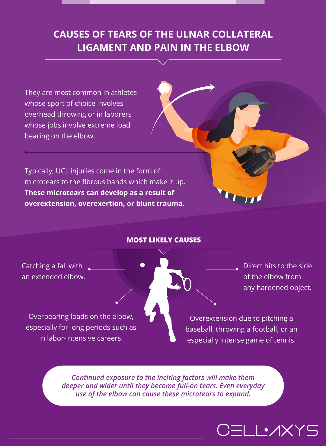
 UCL injuries are fairly common, especially in athletes whose sport of choice involves overhead throwing or in laborers whose jobs involve extreme load bearing on the elbow.
Typically, UCL injuries come in the form of microtears to the fibrous bands which make it up. These microtears can develop as a result of overextension, overexertion, or blunt trauma. Some of the most likely causes are:
- Catching a fall with an extended elbow.
- Overbearing loads on the elbow, especially for long periods such as in labor-intensive careers.
- Direct hits to the side of the elbow from any hardened object.
- Overextension due to pitching a baseball, throwing a football, or an especially intense game of tennis.
Once microtears have developed, continued exposure to the inciting factors will make them deeper and wider until they become full-on tears. Even everyday use of the elbow can cause these microtears to expand.
Once they are present, the only way to combat their progression is to rest. If these injuries develop into something larger, an individual will begin to experience a wide range of symptoms.
Symptoms and Signs of Tears of the Ulnar Collateral Ligament of the Elbow
As UCL injuries develop, an individual will begin to experience several symptoms that could indicate more severe consequences down the road.
If you believe you’ve suffered an injury to the UCL, watch out for these common symptoms:
- Pain
- Swelling
- Instability
- Reduced load-bearing capability
- Reduced range of motion
- Tenderness at the sides of the elbow
In the initial stages of an injury, these symptoms will express themselves slowly. An individual may be able to continue with their daily activity, but strenuous physical exertion will present limitations. Over time, these symptoms will develop into a chronic problem. If these symptoms have begun to present themselves, it may be time to seek professional assistance.
Diagnosing Tears of the Ulnar Collateral Ligament
Diagnosis for a tear in the UCL tissue comes in 3 parts:
- Medical history evaluation
- Physical examination
- Medical imaging
Doctors will begin by examining the patient’s medical history. Have there been any signs of trouble relating to the elbow up until now? Has the patient suffered any trauma to the elbow or surrounding regions in the past? Have previous examinations shown peculiarities within the structures of the elbow?
A doctor must take all of these issues into account before they can pinpoint the precise reason for a patient’s elbow discomfort.
Once the doctor has established a UCL tear as a probable cause of the patient’s pain, they will perform a physical examination. There are various tests a doctor can conduct to rule out other possibilities, but the most common is fairly simple.
The doctor will simply place a small amount of pressure on the side of the elbow, right where the UCL should be. Next, they will slowly push and pull the lower arm inwards and outwards to recreate the symptoms a patient has been feeling. Depending on how much pressure needs to be applied to the UCL and how much pain the test creates, a doctor will accurately be able to measure the progression of the tear.
Finally, with all other tests concluded, the doctor will choose one, or several, medical imaging tests to get an inside view of the elbow and its tissues. The most common and detailed test if a UCL tear is suspected is a magnetic resonance imaging (MRI) scan. This not only provides doctors with a view of the UCL but also the state of the other soft tissues within the elbow.
Degeneration of any of these soft tissues may be another inciting factor for a patient’s pain, instability, swelling, and other symptoms. If the UCL is torn, this will lead to malalignment of the bones within the elbow which may lead to the breaking down of the protective patches of cartilage that line the rims of each bone.
If these tests prove conclusive, the doctor will move on to examining the patient’s functional goals and try to define a clear path to achieving those goals through conventional treatments.
Conventional Treatments for Tears of the Ulnar Collateral Ligament and Pain in the Elbow
Conventional treatments for UCL tears are those treatments that have been most studied and therefore are the most typically recommended by doctors. These treatments include physical therapy, medication, steroidal injections, and, in advanced cases, surgery.
Physical Therapy
Physical therapies aim to restore function and reduce pain via physical methods. Treatments such as massage, guided exercise, stretching, rest, compression, and hot/cold therapy all fall into this category.
The benefit of physical therapies for most patients is the fact that they can be done at home, on the go, or with trained professionals. All levels of physical therapy have been shown to help treat pain and reduce flare-ups of symptoms in some way.
Medication
Most patients will have already tried several over-the-counter medications before consulting a doctor for further assistance. Ibuprofen, naproxen, and acetaminophen are the generic versions of popular name brands that help to reduce inflammation and increase blood flow. These medications aim to stop the painful constriction of the nerves within the elbow after a UCL tear and help patients regain a bit of their mobility without having to move on to more advanced treatments.
If the progression of the tear has become too severe for these medications to have any helpful effect, doctors may move on to prescribing medications aimed at blocking the brain’s ability to interpret pain signals. It should be noted that none of these medications will help to treat the actual cause of the pain, but simply alleviate the symptoms of the tear.
Steroidal Injections
Another popular treatment for pain related to the UCL is steroidal injections. Like over-the-counter medications, these treatments are aimed at reducing swelling and increasing blood flow within the elbow.
While these treatments are effective at counteracting the body’s natural defense mechanisms, in recent years these treatments have come under fire for their degenerative effects. It has been shown that prolonged use of steroids can lead to adverse effects that progress the issues they are trying to combat.
Surgery
Surgery is a last resort for only the most advanced cases of UCL injury, and with good reason. These surgeries rarely work out to the benefit of the patient. While they may reduce the pain a patient feels, the side effects – a reduced range of motion, long recovery periods, bouts of physical therapy and medication – may simply not be worth it for a patient.
If surgery is ultimately the patient’s choice, it typically involves a procedure known as Tommy John surgery, named for the first person to ever receive the treatment. The procedure is applied when the ligament has completely torn off the bone. By making an incision on the inside of the elbow and reattaching the ligament to the bone using sutures, the ligament may heal back into place.
If the UCL is completely degenerated, reconstruction surgery may take place. By extracting and grafting a portion of the ligament in the wrist (palmaris longus), the new tissue may become a suitable replacement for the degenerated UCL.
Though conventional treatments are the most commonly recommended, they are not always the right choice given a patient’s lifestyle and functional goals. If these treatments simply fail to help a patient after they’ve already been performed, the patient will want to seek another route.
Thankfully, there are some alternative treatments at the disposal for patients who fall into this category. One such treatment, regenerative therapy, is quickly making a name for itself due to its relatively easy application and noteworthy results.
Orthobiologic Treatments for Tears of the Ulnar Collateral Ligament and Pain in the Elbow
Unlike conventional treatments, regenerative therapies or orthobiologic methods are focused on treating the cause of the pain rather than the pain itself. As the name implies, they are treatments that attempt to restore damaged tissues.
At CELLAXYS, we use platelet-rich plasma (PRP therapy) or cell-based or stem-cell therapies to heal ulnar collateral ligament tears of the elbow.
PRP Therapy
PRP therapy involves extracting platelets from a patient’s blood and reinjecting them to the injury site. Platelets are the body’s natural first-line defenses against injuries. They form clots that stop bleeding and the progression of further injury. They release 10 Growth Factors that stimulate tissue growth and send chemical signals to attract healing cells in the blood.
Platelets also form fibrin, a web-like sticky matter that supports new tissue building in the injured area. As the affected receive repair tissues, they heal faster and better.
PRP therapy is a popular orthobiologic treatment for sports, spine, and orthopedic injuries. The procedure is usually completed within 45 minutes.
Cell-Based Therapies
Also known as stem-cell therapies, these treatments focus on your body’s autologous tissues and cells. They harvest healthy tissues from your body and reinject them into the affected areas. Depending on your tear, the doctor may recommend you one of the two types of cell-based therapies:
- Minimally Manipulated Adipose Tissue Transplant (MMAT). This method harvests healthy cells from your adipose tissues, processes them, and reinjects them into your affected elbow area. If you need MMAT in multiple areas, your doctor can perform all of them in the same procedure.
- Bone Marrow Concentrate (BMAC). This procedure harvests cells from your bone marrow and injects them into the injury site.
These procedures are performed under anesthesia. They take about 1.5-2 hours to complete and are outpatient procedures, which means you will go home after the procedure.
Sources
Footnotes
- Podesta L, Crow SA, Volkmer D, Bert T, Yocum LA. Treatment of partial ulnar collateral ligament tears in the elbow with platelet-rich plasma. The American journal of sports medicine. 2013;41(7):1689-94.
- Timmerman LA, Andrews JR. Histology and arthroscopic anatomy of the ulnar collateral ligament of the elbow. The American Journal of Sports Medicine. 1994;22(5):667-73.
- Kuroda S, SAKAMAKI K. Ulnar collateral ligament tears of the elbow joint. Clinical Orthopaedics and Related Research (1976-2007). 1986;208:266-71.
- Erickson BJ, Bach Jr BR, Verma NN, Bush-Joseph CA, Romeo AA. Treatment of ulnar collateral ligament tears of the elbow: is repair a viable option?. Orthopaedic Journal of Sports Medicine. 2017;5(1):2325967116682211.
- Azar FM, Andrews JR, Wilk KE, Groh D. Operative treatment of ulnar collateral ligament injuries of the elbow in athletes. The American Journal of Sports Medicine. 2000;28(1):16-23.
References
- Tommy John Surgery (Ulnar Collateral Ligament Reconstruction). Johns Hopkins Medicine. Accessed 2/26/2024.
- Ulnar Collateral Ligament Elbow Injury Diagnosis and Treatment. Penn Medicine. Accessed 2/26/2024.
- Ulnar Collateral Ligament (UCL) Injuries. Cleveland Clinic. Accessed 2/26/2024.
- Corticosteroid Injections of Joints and Soft Tissues. Medscape. Accessed 2/26/2024.
CELLAXYS does not offer Stem Cell Therapy as a cure for any medical condition. No statements or treatments presented by Cellaxys have been evaluated or approved by the Food and Drug Administration (FDA). This site contains no medical advice. All statements and opinions are provided for educational and informational purposes only.
Dr Pouya Mohajer
Author
Pouya Mohajer, M.D. is the Director of Spine and Interventional Medicine for CELLAXYS: Age, Regenerative, and Interventional Medicine Centers. He has over 20 years of experience in pain management, perioperative medicine, and anesthesiology. Dr. Mohajer founded and is the Medical Director of Southern Nevada Pain Specialists and PRIMMED Clinics. He has dedicated his career to surgical innovation and scientific advancement. More about the doctor on this page.
Dr Pejman Bady
Contributor
Dr. Pejman Bady began his career over 20 years ago in Family/Emergency Medicine, working in fast-paced emergency departments in Nevada and Kansas. He has served the people of Las Vegas as a physician for over two decades. Throughout this time, he has been met with much acclaim and is now the head of Emergency Medical Services in Nye County, Nevada. More about the doctor on this page.


