Published on: October 19, 2019 | Updated on: August 29, 2024
Each of our joints is supported by a vast network of soft tissues. One of the most important of these is the tendons. Tendons serve the function of attaching muscle to bone and are a crucial part of most movements throughout the body.
Tendons can become injured or damaged, leading to tendonitis. In the beginning stages, tendonitis is hard to identify but easy to treat. If left untreated, it can become more serious and may even require surgery.
The posterior tibial tendon, located in the ankle, is one of the most important tendons of the foot and ankle, offering structural support and a range of motion for the foot. This tendon is particularly susceptible to developing tendonitis due to its frequent use.
Do you have Posterior Tibial Tendonitis?
Fill out the form below to schedule your FREE virtual consultation
Anatomy of the Ankle
The bones which make up the shin are the tibia and fibula. These bones are in the top part of the ankle joint, and they are connected to the talus inside the foot.
Tendons serve the function of joining muscles to bones. The tibial tendon begins in the foot and extends up into the shin, attaching bones in the foot to calf muscles. The posterior tibial tendon is a major part of arch support and is used in almost all functions of the foot.
Tendonitis (sometimes spelled as tendinitis) occurs when a tendon is irritated, inflamed, or somehow damaged. Some of the common symptoms of tendonitis are:
- Swelling around the tendon (ankle)
- Tenderness of the affected area
- Pain which can feel like a sharp, shooting sensation or a dull ache
- Instability in the joint
- Difficulty using the joint
A traumatic injury could cause tendonitis to occur, but the most common cause is overuse and repetitive motion. Individuals who have a job or hobbies which force them to use the tibial tendon in the same way repetitively, such as a biker or piano player, are at a higher risk of developing posterior tibial tendonitis. Soft tissues in the body also degenerate over time, and this gradual degeneration can cause problems such as arthritis or tendonitis.
Degeneration of the posterior tibial tendon can lead to flat feet, so it is imperative that an injury is spotted and diagnosed in the beginning stages. Some of the damage that this condition incurs can be permanent if it is not diagnosed promptly.
When to See a Doctor
Tendonitis can feel like a dull ache or pain, or it can develop into severe pain that interferes with daily activities. The nature of the injury will determine whether or not it is necessary to visit a doctor.
Many small tears to the tendon can heal on their own by simply resting the joint. If it does not go away in a matter of days, however, and begins to impede daily life, consultation with a doctor may be necessary.
Doctors will inquire about a patient’s activity level, job, and hobbies which may contribute to injury, family history, and more. It is important to be completely honest with them about this information.
The more information a doctor has about an injury, the more likely they are to diagnose it correctly. The degree of injury can typically be determined with a series of tests and imaging technologies. Diagnosing tendonitis typically involves:
- Range of motion tests: doctors will perform certain tests involving the foot, ankle, and leg to determine the source of pain. Pain during certain exercises and the severity of the pain can be indicative of posterior tibial tendonitis. They may also check to see in what directions the foot can move and how far. Tendon degeneration can sometimes prevent certain motions. A doctor may also look for swelling or tenderness in the ankle and foot.
- MRIs: Magnetic resonance imaging, or MRIs, are useful in determining the structure of soft tissue in a patient. Doing so could give doctors a better idea of the extent of injury or degeneration that may be causing pain.
- X-rays: an X-ray will show the bone structure of the foot, which may be useful in determining the severity of the tendonitis. Structural changes may occur during degeneration and these changes would be detectable using X-ray.
- CT scan: this form of imaging can show some soft tissue as well as bone structure.
- Ultrasound: a much less invasive imaging technology that can produce an image of soft tissue and therefore point out potential abnormalities that may be causing pain.
Each imaging technique has its advantages and disadvantages. Doctors will determine which is best for the patient based on their comfort level, financial flexibility, and suspected cause of pain.
Treating Posterior Tibial Tendonitis

Treatment for soft tissue injuries often begins with simply resting the area of injury. The human body has an incredible capacity to heal after trauma, and if the soft tissue is allowed to rest it will often heal on its own. Added stress will prolong the healing process. Rest is often accompanied by the use of over-the-counter NSAIDs (Non-Steroidal Anti-Inflammatory Drugs) such as Ibuprofen.
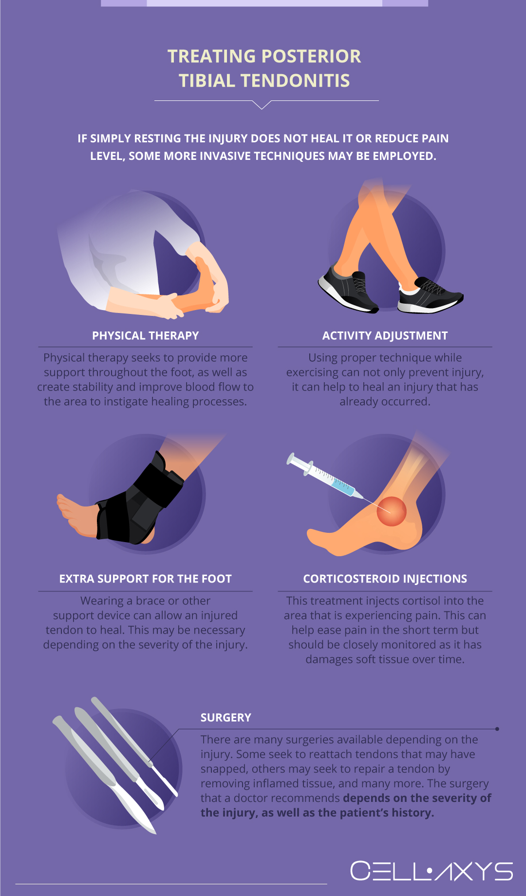
If simply resting the injury does not heal it or reduce pain level, some more invasive techniques may be employed. These include:
- Physical therapy: the goal of physical therapy is generally to improve range of motion and strengthen support muscles. For the posterior tibial tendon, physical therapy seeks to provide more support throughout the foot, as well as create stability and improve blood flow to the area to instigate healing processes.
- Activity adjustment: using proper technique while exercising can not only prevent injury, it can help to heal an injury that has already occurred. Good form during exercise can have a myriad of benefits.
- Extra support for the foot: wearing a brace or other support device can allow an injured tendon to heal. This may be necessary all the time or only during certain activities, depending on the severity of the injury.
- Corticosteroid injections: this form of treatment injects cortisol, a synthetic anti-inflammatory drug, into the area that is experiencing pain. This can help ease pain in the short term but should be closely monitored for long-term use as it has been proven to damage soft tissue over time.
- Surgery: when less invasive methods fail to ease pain and functionality, doctors may turn to surgical solutions. For tendonitis in the ankle, these surgeries can be short and minimally invasive. They can also be intensive procedures that have a long recovery time. There are many surgeries available depending on the injury. Some seek to reattach tendons that may have snapped, others may seek to repair a tendon by removing inflamed tissue, and many more. The surgery that a doctor recommends depends on the severity of the injury, as well as the patient’s history.
Doctors will choose to recommend different treatments based on the specific needs of the patient. Degeneration can not always be avoided but can be managed effectively. Patients who are experiencing ankle pain should consider all of the treatments on the market. Regenerative medicine is a field that may offer a faster recovery time after the injury has occurred.
Regenerative Medicine’s Role in Reducing Recovery Time of Tendonitis
Regenerative medicine offers an option for treating degeneration and soft tissue injury. These treatments are proven to be effective in soft tissue such as tendons. They can be used at any stage in the injury process.
Tibial tendinitis can permanently impair the foot’s structure. Although regenerative medicine is not a cure for these injuries, it can assist in alleviating the discomfort they cause. If the injury or degeneration is not diagnosed until it is more advanced, these therapies can still help by healing the damage that has been done and preventing further injury.
The two types of regenerative therapy that CELLAXYS offers are:
- Cell-based therapies: Also known as stem cell therapies, this form of treatment uses a patient’s own cells or “autologous tissues.” They are extracted from different parts of the patient, processed to become more concentrated, and then injected into the injury site. Depending on the patient’s condition, the doctor may extract healthy cells from the adipose (fat) tissue; the process is called Minimally Manipulated Adipose Tissue (MMAT) transplant, or from the bone marrow; called Bone Marrow Concentrate (BMAC).
- Platelet-rich plasma (PRP) therapy: This form of therapy draws the patient’s blood to isolate platelets from the plasma. The processed platelets are then injected into the injured soft tissue of the patient. Platelets contain proteins and 10 Growth Factors that encourage natural healing processes and the development of healthy tissues. Platelets also attract healing cells from the blood and produce a web-like scaffolding called fibrin to support the growth of healthy tissues. Increasing the number of platelets in the injured area may help heal the injury faster.
Both forms of therapy are outpatient procedures, meaning you can go home right after the process. Cell-based therapies are completed within 1.5 to 2 hours, while PRP takes about 45 minutes. The doctors use a live X-ray or ultrasound to pinpoint the damage’s location. Doing this allows doctors to have as much precision as possible.
They can also be coupled with surgery to aid in the recovery process. Depending on the surgery, this process can take months. Using regenerative medicine could shorten this.
It is still often recommended that patients stick to the original recovery plan after surgery as well, including physical therapy and medication. These therapies offer a solution to help with pain and healing, but a patient’s recovery process is still heavily dependent on the patient’s willingness to commit to recovery.
Sources
Footnotes
- Alvarez RG, Marini A, Schmitt C, Saltzman CL. Stage I and II posterior tibial tendon dysfunction treated by a structured nonoperative management protocol: an orthosis and exercise program. Foot & ankle international. 2006;27(1):2-8.
- Chao W, Wapner KL, Lee TH, Adams J, Hecht PJ. Nonoperative management of posterior tibial tendon dysfunction. Foot & ankle international. 1996;17(12):736-41.
- Mendicino SS. Posterior tibial tendon dysfunction: diagnosis, evaluation, and treatment. Clinics in podiatric medicine and surgery. 2000;17(1):33-54.
- Myerson MS, Corrigan J. Treatment of posterior tibial tendon dysfunction with flexor digitorum longus tendon transfer and calcaneal osteotomy. Orthopedics. 1996;19(5):383-8.
References
- Tibialis posterior tendonitis – Top treatments … and what to avoid!. Sports Injury Physio. Accessed 2/25/2024.
- Posterior Tibial Tendon Dysfunction (PTTD). Cleveland Clinic. Accessed 2/25/2024.
- Posterior Tibial Tendonitis Causes, Symptoms & Treatment. Upswing Health. Accessed 2/25/2024.
CELLAXYS does not offer Stem Cell Therapy as a cure for any medical condition. No statements or treatments presented by Cellaxys have been evaluated or approved by the Food and Drug Administration (FDA). This site contains no medical advice. All statements and opinions are provided for educational and informational purposes only.
Dr Pouya Mohajer
Author
Pouya Mohajer, M.D. is the Director of Spine and Interventional Medicine for CELLAXYS: Age, Regenerative, and Interventional Medicine Centers. He has over 20 years of experience in pain management, perioperative medicine, and anesthesiology. Dr. Mohajer founded and is the Medical Director of Southern Nevada Pain Specialists and PRIMMED Clinics. He has dedicated his career to surgical innovation and scientific advancement. More about the doctor on this page.
Dr Pejman Bady
Contributor
Dr. Pejman Bady began his career over 20 years ago in Family/Emergency Medicine, working in fast-paced emergency departments in Nevada and Kansas. He has served the people of Las Vegas as a physician for over two decades. Throughout this time, he has been met with much acclaim and is now the head of Emergency Medical Services in Nye County, Nevada. More about the doctor on this page.


