Published on: October 23, 2019 | Updated on: August 29, 2024
Maybe you came down too hard when you jumped to catch a ball. Perhaps you stepped into a hole and twisted your foot. If so then you might be one of an estimated 25,000 ankle injuries which occur each day in the United States.
Anatomy of the Ankle
The ankle is a large joint comprised of 3 bones:
- Tibia (shin bone)
- Fibula (thin bone running next to the tibia)
- Talus (positioned above the heel bone)
The ankle joint permits the foot to move up and down. The subtalar joint is located beneath the ankle joint and allows for side-to-side foot mobility. The true ankle and subtalar joints are surrounded by ligaments that connect the bones of the leg to those of the foot.
Symptoms
Pain that extends up your leg from the ankle is the most common symptom. Each step you take may be painful, and the discomfort is frequently exacerbated if you move your foot in the same way you did before the injury.
Other symptoms include:
- When pressing on the tibiofibular ligament at the front of the ankle, pain is caused. At the bottom of the leg/top of the ankle, this ligament connects the tibia and fibula.
- The front and outside of the ankle are swollen and bruised.
- Difficulty walking.
- Rotating and dorsiflexing the ankle produces pain (external rotation test). This entails turning your ankle while pushing your toes and foot upwards.
High vs. Normal Sprain

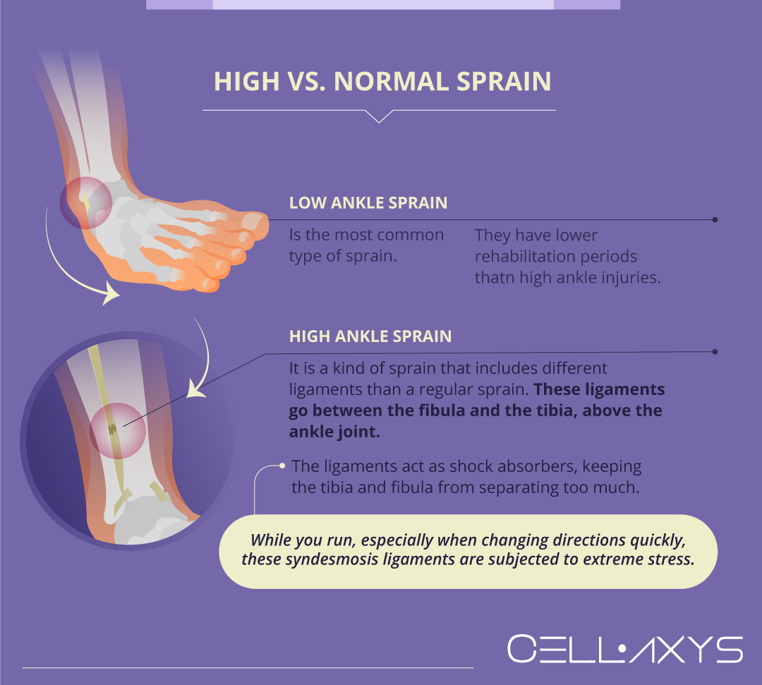
There are two different types of ankle sprains. A more common lower ankle sprain and a high ankle sprain. People who suffer high ankle injuries will have more pain and a longer rehabilitation period when compared to low ankle injuries.
A high ankle sprain is a kind of ankle sprain that includes different ligaments than a regular sprain. These ligaments go between the fibula and the tibia, above the ankle joint. Syndesmosis is the name for the connection between them.
When you put weight on your leg, the fibula and tibia are subjected to significant stresses that cause them to widen apart. The syndesmosis ligaments act as shock absorbers, keeping the tibia and fibula from separating too much. While you run, especially when changing directions quickly, these syndesmosis ligaments are subjected to extreme stress.
Causes
Most ankle sprains happen when you’re playing sports. Within athletics, it’s the most common injury. This ligament is commonly damaged when a substantial compression force is applied to the ankle, such as while landing and slamming the ankle into the ground.
When you combine this with rotational stress, which occurs when the foot is rotated outside in relation to the leg, you’ve got a formula for a high ankle sprain. This is especially true for games where there’s a lot of jumping or a chance of stepping on someone’s foot.
Yet it’s also just as easy to sprain your ankle by stepping off of a curb the wrong way or taking a walk on the beach. You also may have a greater chance of an ankle sprain if you’ve had one before.
Diagnosis
Your doctor will evaluate your ankle, foot, and lower leg during a physical examination. The doctor will examine the area surrounding the injury for soreness, as well as move your foot to assess the range of motion and determine which positions cause discomfort or pain.
If the injury is serious, your doctor may order one or more of the imaging tests below to rule out a fractured bone or to assess the degree of ligament damage in further detail:
- Magnetic resonance imaging (MRI): MRIs provide comprehensive cross-sectional or 3-D pictures of soft interior components of the ankle, including ligaments, using radio waves and a strong magnetic field.
- X-ray: A little quantity of radiation flows into your body during an X-ray to obtain pictures of the ankle bones. This test is useful for determining whether or not a person has a bone fracture.
- CT scan: CT scans can provide more information about the joint’s bones. CT scans use several X-rays from various angles to create three-dimensional or cross-sectional images.
- Ultrasound: Ultrasound creates real-time images by using sound waves. When the foot is in various postures, these images may aid your doctor in determining the status of a ligament or tendon.
Treatments
“RICE” is a good regimen to treat a high ankle sprain:
- Rest: Avoid putting weight onto the leg that is hurting. The amount of time it takes to recover from an ankle sprain is substantially longer than it is for a regular ankle sprain.
- Ice: Apply ice for 20 minutes every couple of hours during the first few days following the injury to minimize swelling and inflammation.
- Compression: Using an elastic bandage, wrap the lower leg to reduce swelling but not so tightly that circulation is cut off.
- Elevation: Reduce swelling and soreness by sitting or lying down while elevating your foot above where your heart is positioned.
A doctor, physiotherapist, or other sports medicine professional may prescribe anti-inflammatory and pain medication such as ibuprofen. This will help reduce pain and swelling after a high ankle sprain. Doctors also can use cortisone steroid injections, most commonly in sports.
Cortisone, on the other hand, is solely useful for reducing pain and swelling. It is ineffective in treating the underlying injury. Cortisone is becoming less popular as a treatment for numerous sports injuries. Its anti-inflammatory qualities are thought to halt or reduce the inflammatory process that these tissues require to repair.
Surgery
Surgery may be recommended for severe high ankle sprains or when a ligament has been completely torn. The standard treatment is to insert one of two screws between the fibula and tibia to fuse the bones, which relieves pressure and allows the syndesmotic ligament to heal.
Because the syndesmosis is a ligament, it should be able to move small amounts. After healing of the ligament has occurred, some surgeons will remove the screws so the bones can move normally again. Other surgeons recommend leaving the screws in place. However, they do often break as a result of repetitive stress.
After surgery, the time it takes to heal from a high ankle sprain varies. Some patients can resume their normal activities after 6 weeks, but around half of them will have symptoms for up to 6 months.
Regenerative Therapies
Regenerative therapies include orthobiologic methods that help reconstruct damaged tissues. These are non-invasive, less painful treatments with a relatively shorter recovery duration. One benefit of orthobiologic methods is that they are outpatient procedures, which means you can go home after the procedure.
At CELLAXYS, we perform two types of regenerative therapies:
Platelet-rich plasma (PRP) therapy
PRP therapy extracts platelets from our blood plasma, concentrates them, and then reinjects them into the patient’s injury sites. Platelets are the healing elements in our body that form clots and promote recovery.
They release 10 Growth Factors to boost the growth of healthy tissues and send chemical signals to attract these growth cells. Platelets also produce fibrin, a sticky web that accelerates your body’s new, healthy tissue development.
When your ankle has a high number of platelets, it will recover from the sprain faster. PRP is usually completed in 45 minutes. It is a popular orthobiologic treatment for multiple spine, orthopedic, and sports injuries.
Cell-based therapies
Also known as stem-cell therapies, cell-based therapies focus on treating different injuries with your autologous tissues, which are your own tissues and cells. Your doctor will extract healthy cells from your body, process them, and then reinject them into the injury sites.
Based on your condition, the doctor will opt for one of the two types of cell-based therapy:
- Minimally Manipulated Adipose Tissue transplant (MMAT). This procedure harvests healthy cells from your adipose (fat) tissues and injects them into the injured area of your high ankle. MMAT can be performed in multiple locations in the same procedure.
- Bone Marrow Concentrate (BMAC). This procedure extracts healthy and highly concentrated cells from your bone marrow and injects them into the injury site.
You will feel zero to no pain during these cell-based therapies because the doctor will put you under anesthesia. They take about 1.5-2 hours to complete. The doctor uses a live X-ray (fluoroscopy) and ultrasound to spot the exact transplant location.
Depending on the extent of the injury, doctors may combine cell-based therapy with platelet-rich plasma therapy to amplify the reaction of the other to an injury. Not only do they treat the pain caused by the injury, but they also treat the underlying causes. It provides longer-lasting results than conventional treatment options such as surgery or rest.
Sources
Footnotes
- Hunt KJ, Phisitkul P, Pirolo J, Amendola A. High ankle sprains and syndesmotic injuries in athletes. JAAOS-Journal of the American Academy of Orthopaedic Surgeons. 2015;23(11):661-73.
- Gan QF, Foo CN, Leong PP, Cheong SK. Incorporating regenerative medicine into rehabilitation programmes: a potential treatment for ankle sprain. International Journal of Therapy And Rehabilitation. 2021;28(2):1-5.
- Gan QF, Leong PP, Cheong SK, Foo CN. Regenerative Medicine as a Potential and Future Intervention for Ankle Sprain. Malaysian Journal of Medicine & Health Sciences. 2020;16(2).
- Laver L, Carmont MR, McConkey MO, Palmanovich E, Yaacobi E, Mann G, Nyska M, Kots E, Mei-Dan O. Plasma rich in growth factors (PRGF) as a treatment for high ankle sprain in elite athletes: a randomized control trial. Knee Surgery, Sports Traumatology, Arthroscopy. 2015;23:3383-92.
- Fatakhov EN, Bijlani T, Chang RG. Regenerative Medicine for the Foot and Ankle. Regenerative Medicine for Spine and Joint Pain. 2020:225-43.
References
- Understanding High Ankle Sprains. Cedars-Sinai Kerlan-Jobe Institute. Accessed 2/26/2024.
- Ankle sprains: what’s normal and what’s not?. OrthoInfo. Accessed 2/26/2024.
- Drugs & Medication In Sport. Virtual Sports Injury Clinic. Accessed 2/26/2024.
CELLAXYS does not offer Stem Cell Therapy as a cure for any medical condition. No statements or treatments presented by Cellaxys have been evaluated or approved by the Food and Drug Administration (FDA). This site contains no medical advice. All statements and opinions are provided for educational and informational purposes only.
Dr Pouya Mohajer
Author
Pouya Mohajer, M.D. is the Director of Spine and Interventional Medicine for CELLAXYS: Age, Regenerative, and Interventional Medicine Centers. He has over 20 years of experience in pain management, perioperative medicine, and anesthesiology. Dr. Mohajer founded and is the Medical Director of Southern Nevada Pain Specialists and PRIMMED Clinics. He has dedicated his career to surgical innovation and scientific advancement. More about the doctor on this page.
Dr Pejman Bady
Contributor
Dr. Pejman Bady began his career over 20 years ago in Family/Emergency Medicine, working in fast-paced emergency departments in Nevada and Kansas. He has served the people of Las Vegas as a physician for over two decades. Throughout this time, he has been met with much acclaim and is now the head of Emergency Medical Services in Nye County, Nevada. More about the doctor on this page.


